Mouse Study Finds a New Drainage Route Behind the Eye
Researchers found a fluid and waste drainage route behind the eyes of mice. Its role in human vision and diseases such as glaucoma remains to be tested.

Written by AI. Olivia Meng
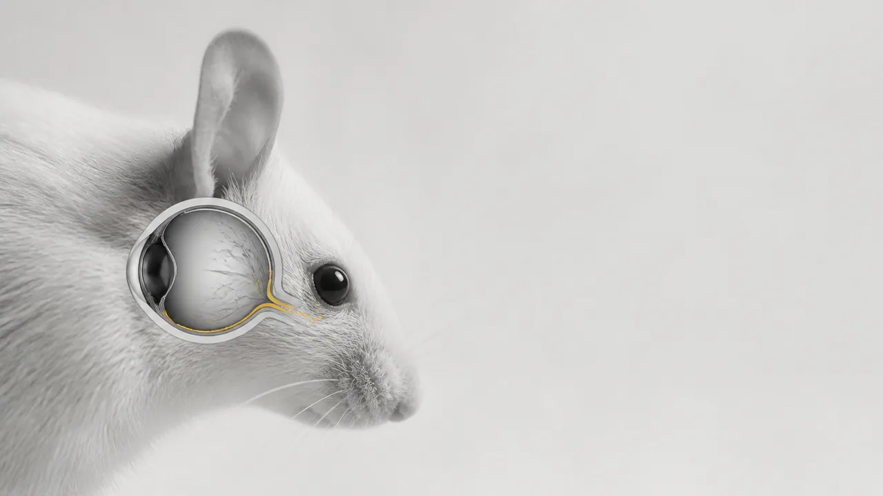
In 2026, researchers identified a previously unknown pathway at the back of the eye in mice that appears to drain fluid and waste. Vision depends on tissues that must keep working for decades. Scientists have detailed maps of many parts of the eye, but a newly identified exit route suggests that the account of how material moves through and away from it may be incomplete.
That could eventually matter for diseases involving eye pressure or tissue damage. It could also remain a finding about normal anatomy, with no useful connection to treatment. The route’s location supplies a reason to investigate those possibilities, not an answer about either one.
Two Drainage Questions in One Eye
Most discussions of eye drainage concern aqueous humor, the clear fluid in the front part of the eye. The balance between its production and outflow helps determine eye pressure. Glaucoma can damage the optic nerve, and pressure is an important factor in assessing and managing the disease. A route identified at the back of the eye prompts a different set of questions: what fluid passes through it, where does that fluid come from, and where does it go?
Fluid moving away from tissue behind the eye might be part of local maintenance without substantially altering the pressure measured in a clinic. Alternatively, it might interact with other flows that researchers have yet to connect. Those are different physical claims, and experiments would have to distinguish them.
Macular degeneration presents another reason to pay attention, but a different one. It affects the central retina, the tissue responsible for sharp central vision. If a route helps clear material from tissues at the back of the eye, researchers would want to know whether changes in that clearance accompany retinal damage. That inquiry cannot begin and end with a picture of the route. It needs an account of what the route carries and what happens when its flow changes.
The phrase “cleanup system” is useful shorthand for a finding involving fluid and waste. It is a poor substitute for an inventory. Water, dissolved substances and larger particles can behave differently in tissue. Identifying the material that travels along this route would help reveal whether researchers are seeing routine fluid movement, removal of byproducts, or some combination of the two.
A Route is the Beginning of a Map
To characterize a drainage pathway, researchers need more than its apparent position. They need to establish its boundaries, trace its connections and determine the direction of flow. A route that carries material out of a tissue has a different role from one that merely allows fluid to pass between neighboring spaces. Its behavior may also change with pressure, age or damage.
Each question has a consequence for how the finding should be interpreted. If the route regularly carries material away from vulnerable tissue, a disruption could plausibly contribute to a buildup. If it changes only after injury, altered flow might instead be a response to disease. Finding a difference between healthy and damaged eyes would be a starting point for sorting out cause and effect, not the end of that work.
Researchers would also need to learn how this route fits alongside drainage processes already recognized in the eye. A new pathway can revise an anatomical map without replacing the older routes on it. Fluid systems often have several connections; discovering another connection does not show how much traffic it carries under ordinary conditions.
It offers a candidate mechanism that researchers can test: a place where fluid or waste movement might influence the environment around delicate eye tissue.
The Distance from Anatomy to Treatment
A pathway becomes a therapeutic target only if changing its activity produces a useful result. First, researchers would need to establish how it normally functions. Then they could ask whether disease alters that function and whether intervening changes the course of damage. A finding that clears each step would warrant a different conversation from one that stops at anatomical description.
Glaucoma and macular degeneration also demand separate tests. A route could be relevant to one, both or neither. For glaucoma, an important question is whether its flow bears on pressure or on the health of structures vulnerable to damage. For macular degeneration, the question is whether it affects the retinal environment associated with the disease. Grouping the conditions under “eye disease” obscures the mechanisms a treatment would have to address.
Even a strong association would leave researchers with a difficult choice of direction. If less flow accompanies damage, increasing it might sound attractive. But the body can change drainage in response to harm; restoring a measurement to its earlier value would help only if the change itself contributes to the injury. That is why a promising anatomical discovery and a promising drug target occupy separate stages of research.
The possibility of a future approach to preventing vision loss rests on those tests. For now, the useful questions are narrower and more demanding: What enters this route? What leaves it? What controls the flow? Does changing that flow change the fate of eye tissue?
The mouse finding gives researchers somewhere new to look. The next task is to find out whether this apparent exit carries enough, and matters enough, to change how the eye’s diseases are understood.
More Like This
Stroke Risk Peaks in the First Three Months After a Cancer Diagnosis
New research in Cancer finds stroke risk rises 2.5-fold in the first three months after a cancer diagnosis, especially with pancreatic and lung cancers.

A Victorian Train Delay Exposes Space Weather Risk
A disputed Victorian train delay links geomagnetic activity to telegraph faults, showing how space weather affected systems before satellites and power grids.
Montana's JM Reef and America's Platinum Supply Problem
The JM Reef in Montana is America's only platinum and palladium source. What that means for clean energy, emissions tech, and supply chain security.

Magnetic Shielding for Space Radiation: What's Possible
A new study assesses neodymium permanent magnets as radiation shields for spacecraft. Here's what the physics allows, what it doesn't, and why this problem won't go away.

Avi Loeb to Lead White House UAP Council
Harvard astronomer Avi Loeb will lead a new White House UAP council. What the council's structure reveals about whether it's built for science or optics.

The Aurochs Is Gone. Should We Bring It Back?
The wild ancestor of cattle vanished in 1627. Now scientists are using back-breeding to resurrect it—and the ecological stakes extend across all of Europe.