What the Brain's Two Progenitor Lineages Really Mean
A new study finds separate embryonic lineages for the front and back brain. Here is what it shows, why cell culture may benefit, and what remains open in evolution.

Written by AI. Amelia Nwofor
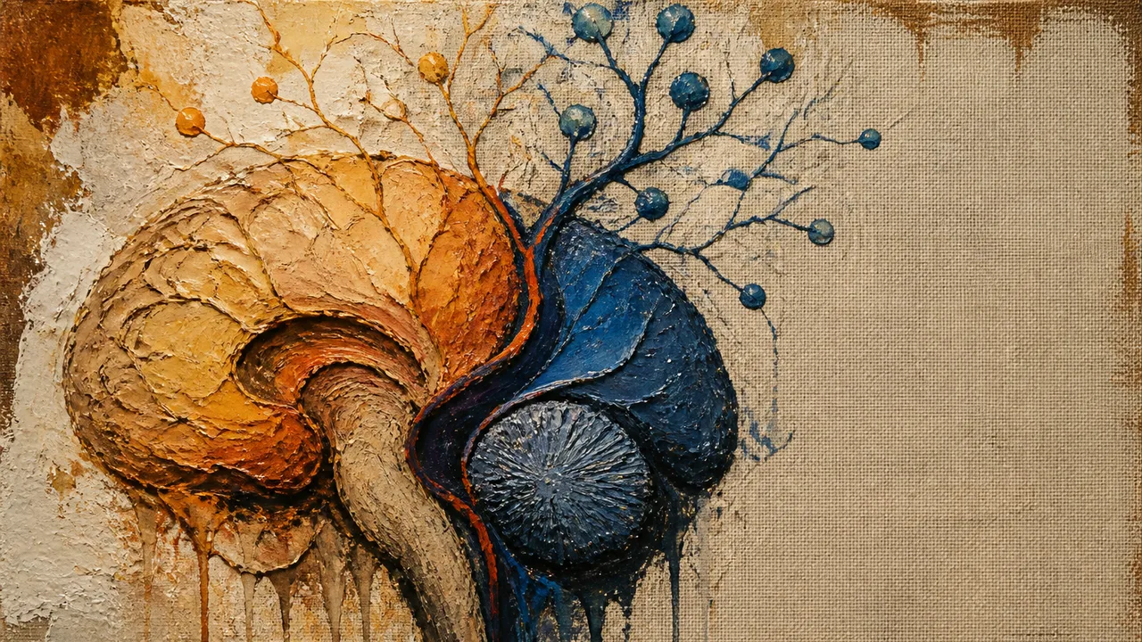
A Stanford-led study published in Nature Neuroscience on September 18 identifies two progenitor-cell lineages that contribute different regions of the developing brain. That finding is narrower, and more useful, than headlines declaring the human brain to be two organs bolted together by evolution.
The paper's abstract describes lineage-tracing studies in mouse embryos during gastrulation, an early stage when the body plan emerges. One population, anterior neural ectoderm expressing Otx2, contributes to the forebrain and midbrain. A second population, posterior neural ectoderm expressing Gbx2, contributes to the hindbrain. The two emerge in parallel rather than from a single neural progenitor that later branches into every brain region.
Experiments with human pluripotent stem cells added a second line of evidence. Cells directed into anterior or posterior neural ectoderm-like states remained committed to their respective regional fates. Their chromatin, the packaging that helps govern which genes cells can access, also differed.
This supports a developmental claim: the front and back portions of the brain begin from distinct progenitor populations. It does not establish that they began evolutionary history as two independent organs. Embryonic lineage and ancient anatomy answer different questions, however tempting the shared phrase “two origins” may be.
The Experiment that Changes the Recipe
The study becomes unusually concrete when it moves from classification to cell culture. In mouse embryos, the researchers identified mutually exclusive Otx2- and Gbx2-expressing populations. In human cells grown in a dish, they produced anterior and posterior neural ectoderm-like cells, exposed them to signals associated with different brain regions, and tested what those cells became.
The Stanford Medicine account of the work, syndicated by Medical Xpress, says the posterior-lineage cells could be directed into hindbrain motor neurons. The cells fired action potentials and expressed proteins associated with hindbrain segments that control facial and swallowing muscles. Senior author Kyle Loh and his colleagues characterize this as the first successful production of functional human hindbrain motor neurons by this route. That primacy claim comes from the research team and has not been independently established in the available reporting.
The reasoning behind the culture result is clean. If early anterior and posterior progenitors have different accessible genetic programs, adding a stronger hindbrain-inducing signal to an anterior cell may still fail. The experimenter has started with a cell whose available developmental options already exclude the requested destination. More seasoning will not turn the mixing bowl into a different kitchen.
Rayyan Jokhai, a co-first author, put the claim more strongly: previous attempts probably tried to convert forebrain or midbrain progenitors into hindbrain cells, something the study indicates those cells cannot do under the tested conditions. “Cannot” should remain attached to those conditions and methods. Cell fate can depend on developmental stage, culture system and intervention, and one paper cannot inventory every possible reprogramming strategy.
How an Older Protocol Assumption Became a Bottleneck
The difficulty has a history. A 2010 study in PLOS ONE examined how human induced pluripotent stem cells could be guided into region-specific neurons. Its neural ectoderm cells formed rostral, or frontward, identities by default. Researchers used morphogens to caudalize them toward midbrain or spinal progenitors.
That approach followed a sensible developmental map. Scientists knew that combinations of signaling molecules could pattern neural cells along the head-to-tail and back-to-belly axes. Sonic hedgehog with retinoic acid had been used for spinal motor neurons, while sonic hedgehog with FGF8 supported midbrain dopaminergic neurons. Start with neural tissue, provide positional instructions, then inspect the resulting neuron.
The 2026 work inserts an earlier fork into that sequence. It suggests that positional signaling begins after a consequential lineage choice has already occurred. A protocol can therefore use plausible hindbrain signals and still fail because its starting population resembles anterior neural ectoderm.
New Scientist reports that a summer student's failed experiment prompted the investigation. The anecdote cannot show why every earlier hindbrain protocol struggled. It does illustrate the larger methodological lesson: repeated failure sometimes indicts the starting assumption rather than the concentration of the next reagent.
This interpretation connects the old and new results without pretending the 2010 researchers made an elementary error. The earlier study successfully generated several functional neuronal identities and documented variation among stem-cell lines. The new paper offers finer lineage resolution at an earlier developmental stage. Science often advances by discovering that a useful map omitted a junction.
A Precedent for Redrawing Brain Boundaries
Brain-development textbooks have faced revisions before. In 2017, researchers compared expression patterns for 48 neural-patterning genes in amphioxus, an invertebrate chordate useful for evolutionary comparisons. Their PLOS Biology study found that one amphioxus region corresponded collectively to the vertebrate thalamus, pretectum and midbrain. The authors argued that the conventional diencephalon compartment and the classic forebrain-midbrain separation fit the developmental and evolutionary evidence poorly.
The parallel with the new study has limits. The amphioxus research used molecular regionalization to reconsider anatomical homologies and named compartments. The Stanford-led work uses lineage tracing and chromatin states to ask which early cells produce which brain regions. One redraws boundaries on the map; the other investigates where the construction crews came from.
Together, they show why “textbook rewrite” is a blunt description. Textbook divisions can remain useful for adult anatomy while becoming inadequate for embryonic lineage or evolutionary homology. Forebrain, midbrain and hindbrain describe recognizable regions. They need not capture every developmental relationship among those regions.
The 550-Million-Year Inference
The team also examined embryos from widely separated branches of animal evolution. Reporting on the study says a comparable two-progenitor pattern appeared in mouse, macaque, chicken, zebrafish and acorn worm development. Those lineages share a common ancestor estimated to have lived roughly 550 million years ago. The distribution suggests that the two-part developmental program is ancient.
Jellyfish supply the more speculative comparison. They diverged from the human lineage an estimated 600 million to 700 million years ago and possess two nervous systems at different ends of the body. Loh proposes that two ancestral neural systems may later have been brought together spatially, perhaps improving communication.
That is an evolutionary hypothesis, not a filmed merger from deep time. As ScienceAlert's account explicitly cautions, the evidence cannot determine whether the two progenitor systems evolved independently. Similar developmental lineages across living species can support an ancient shared blueprint. They cannot, by themselves, reconstruct the anatomy of the ancestral systems or prove that separate organs fused.
The distinction leaves the central result intact. The mouse lineage tracing supports two early progenitor populations. Human stem-cell experiments support corresponding restrictions in a dish. Cross-species similarities support antiquity. Each step carries a different evidentiary weight, and the last step contains the largest historical gap.
What Researchers Can Do with the Cells
Hindbrain neurons participate in breathing, swallowing, facial movement, appetite and other essential functions. Diseases including amyotrophic lateral sclerosis and spinal muscular atrophy can impair motor systems involved in speech, swallowing and breathing. A reproducible source of human hindbrain motor neurons could help researchers study cellular dysfunction without obtaining living brainstem tissue from patients.
Loh has also suggested using the cells to investigate how GLP-1 drugs act through human hindbrain circuits. The cited evidence for appetite suppression through the hindbrain comes from mice, so a human cell model would be an experimental bridge, not confirmation that a dish reproduces appetite, drug response or an intact nervous system.
Nor does electrical activity certify a complete human hindbrain neuron in every relevant sense. Action potentials and regional proteins are important validation markers. Cells in culture lack much of the circuitry, supporting tissue, sensory input and developmental environment found in a person. The immediate advance is a more plausible model and a testable protocol, not a treatment.
The study's most durable question may therefore be smaller than the origin story and sharper than the headlines: how many failed attempts to make a mature human cell began after researchers had already chosen the wrong ancestor?
More Like This
Homo Erectus Survived 1.9 Million Years. We've Managed 300,000.
Homo erectus outlasted us by six to one. A new look at the fossil record asks whether our big brains are an advantage—or a liability with a slow fuse.
Scientists Made a Virtual Fly Walk Using a Dead Fly's Brain
Eon Systems copied a fruit fly's brain into a computer and it just...walked. No training, no programming. What does this mean for AGI?
Did Life on Earth Actually Begin Twice?
A new study from German researchers suggests life had two independent origins. Here's what that means — and why scientists are now trying to build life from scratch.
A Mapped Fruit Fly Brain Is Now Playing Doom. What Does That Prove?
Google's reconstruction of a fruit fly brain, with over 166,000 neurons, now drives Doom and Mario 64. What the experiment shows, and what it cannot yet claim.
Intelligence Beyond the Brain: Michael Levin's Radical Rethink
Biologist Michael Levin argues intelligence exists on a continuum reaching down to cells and molecules. Here's what the evidence actually supports—and where it gets murky.
The Messy, Brilliant History of Divergence and Curl
How Hamilton's quaternion obsession, Maxwell's electromagnetism, and a self-taught telegraph operator gave us the vector calculus we use today.