What Spinal Bone Density Can Say About Brain Aging
A 2026 study links lower spinal bone density on chest CT to faster cognitive decline, but it offers a risk signal, not a dementia diagnosis or proof of cause.

Written by AI. Priya Sharma
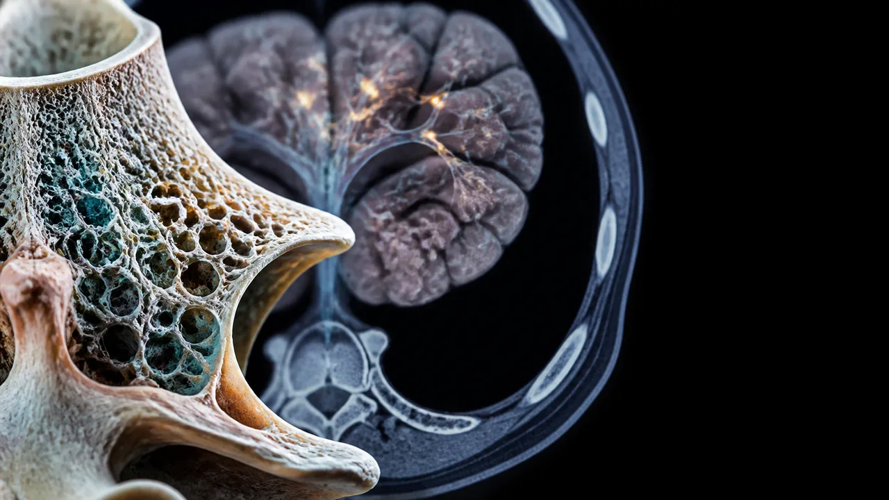
Johns Hopkins researchers extracted spinal bone-density measurements from 2,086 chest CT scans and found that lower density tracked faster cognitive decline in a smaller group followed with brain imaging and cognitive tests.
The result, published in Radiology on September 22, 2026, offers a possible second use for scans originally taken for lung cancer screening, calcium scoring, pulmonary nodule follow-up and other reasons. An existing image could yield information about the thoracic spine without another scan or another radiation exposure.
That possibility is more defensible than calling the measurement an early dementia test. The study found associations, with modest reported effect sizes, in selected participants from an established research cohort. It did not show that bone loss damages the brain, establish an individual diagnostic threshold or test whether acting on the CT-derived result prevents cognitive decline.
What the Researchers Measured
The team conducted a secondary analysis of the Multi-Ethnic Study of Atherosclerosis, or MESA. Participants had no clinically recognized cardiovascular disease when they entered that cohort. For the new analysis, a deep-learning system calculated baseline volumetric bone mineral density, or vBMD, in the thoracic vertebrae from noncontrast chest CT images.
The study team's account says 715 participants with an established CT-derived vBMD also had brain MRI and cognitive testing. Longitudinal white-matter hyperintensity data were available for 408 people, while 405 had longitudinal fractional-anisotropy data. Those shrinking sample sizes are relevant to interpretation: the broadest pool supplied the bone measurements, but the brain-change analyses relied on roughly one-fifth of the 2,086 scanned participants.
White-matter hyperintensities are bright areas on MRI associated with injury and brain aging. Fractional anisotropy measures aspects of white-matter microstructure. Lower baseline vBMD was associated with faster accumulation of hyperintensities in the corpus callosum and steeper fractional-anisotropy decline in the anterior limb of the internal capsule. Both regions contribute to networks involved in executive functions.
The reproduced journal abstract puts numbers on those findings. It reports a coefficient of 12.8% per year for corpus-callosum hyperintensity accumulation among 408 participants, a coefficient of minus 0.048 standard deviations per year for fractional anisotropy, and minus 0.025 standard deviations per year for decline in a global cognitive composite among 639 participants. It also reports a diabetes interaction of 4.99% for white-matter hyperintensity progression.
These coefficients describe average statistical relationships under the study’s models. They do not tell an individual reader how quickly cognition will change, and the supplied abstract does not provide enough detail to translate every coefficient into an absolute personal risk. Statistical significance can identify a pattern without making that pattern large, diagnostic or useful in a clinic.
A Shared Aging Process is the Stronger Explanation
Senior author Shadpour Demehri explicitly rejected a causal bone-to-brain reading. In a statement reproduced by Nautilus, he said the co-occurrence may reflect shared drivers of aging, including insulin resistance, dyslipidemia and menopausal change, rather than a direct effect of bone on the brain.
That explanation fits the study design. Researchers measured bone density at baseline and examined subsequent imaging and cognitive changes, which establishes temporal ordering. They did not assign participants to different bone-density levels or intervene on bone loss. Shared metabolic conditions, hormonal changes or other factors could therefore contribute to both skeletal and neurological decline.
The diabetes interaction strengthens the case for examining shared systemic risk, although it does not identify a mechanism. A reasonable next use of the measurement would be as a prompt for further assessment of skeletal and metabolic health in research or validated clinical pathways. The present study cannot show that such follow-up improves cognition, reduces fractures or changes any other patient outcome.
For someone who has already had a chest CT, the paper supplies no validated instruction to retrieve the image and infer dementia risk from a vertebral value. The algorithm produced a research measurement, and the study did not establish a cutoff separating people who will decline from those who will not. Clinical interpretation would also require evidence that the result adds useful information beyond age, diabetes status, established bone assessment and other known risk factors.
Opportunistic Bone Screening Predates This AI System
Radiologists were investigating spare information in CT scans before deep learning became the headline. A 2020 PLOS ONE study retrospectively examined 414 breast-cancer patients who had both noncontrast chest CT and dual-energy X-ray absorptiometry, or DXA, within three months. The researchers measured attenuation at the L1 vertebra in Hounsfield units and compared it with DXA, the reference method described in that paper.
At a threshold of 90 Hounsfield units, the CT measurement had 54.9% sensitivity and 85.8% specificity for the study’s osteoporosis classification. Raising the threshold to 110 increased sensitivity to 83.9% while lowering specificity to 70.1%. Those trade-offs are screening in miniature: catch more possible cases and generate more false alarms, or miss more cases while making a positive result more selective.
The 2020 paper also cited earlier feasibility work on measuring vertebral attenuation in chest or abdominal CT scans obtained for other indications. Opportunistic bone assessment was therefore an established research program by 2020. The 2026 work changes two elements: deep learning derives a volumetric thoracic-spine measurement, and the outcome under study extends beyond osteoporosis and fracture risk to longitudinal cognition and brain MRI changes.
The comparison also sets boundaries. The earlier study involved a disease-specific cohort of breast-cancer patients and assessed fracture-related outcomes against DXA. The MESA analysis involved a multiethnic cardiovascular research cohort and assessed cognition and white matter. Different populations, measurements and outcomes prevent a direct performance comparison.
Both studies repurposed images and data collected in broader clinical or research settings. Based on their reported designs, neither tested whether routinely returning an opportunistic bone result to patients improved health. The available research also does not establish a standard threshold for using the 2026 algorithm across scanners, hospitals and populations.
A Promising Flag Still Needs a Clinical Destination
Automated analysis could make opportunistic measurement easier to perform at scale. Automation, however, does not answer what clinicians should do with the number, how often it will produce false alarms or whether it improves decisions beyond existing assessments. A biomarker becomes useful when it reliably changes care for the better, not merely when software can extract it.
The immediate contribution of the 2026 study is narrower and scientifically useful. It connects a CT-derived skeletal measure with two different kinds of longitudinal evidence: cognitive performance and regional white-matter change. Agreement across those outcomes makes the association harder to dismiss as an artifact of one cognitive test. Attrition, cohort selection, unverified generalizability and possible residual confounding still limit the inference.
Multiple reports about the finding describe the same Radiology paper and the same author statements. They are coverage of one study rather than replications. Confirmation will require other cohorts, transparent validation of the algorithm, clinically interpretable thresholds and studies that test whether responding to the flag benefits patients.
A chest CT may contain more information than the question that prompted it. The harder task is deciding which extra measurements deserve to leave the research archive and enter a medical record.
More Like This
The Sleep-Deprived Brain: Hidden Costs Revealed
Explore how lack of sleep impacts memory, emotions, decisions, and brain health. Discover why sleep is crucial.

Can Reading for Pleasure Protect Your Brain Health?
A 2026 review links pleasure reading to cognition and wellbeing, but most evidence is observational. Here is what readers can reasonably conclude today.
Taxi Drivers Rarely Die of Alzheimer's
New research shows taxi drivers have unusually low Alzheimer's mortality rates. The reason may say something uncomfortable about GPS and how we all navigate now.

Why Finding Earth's Twin Among Exoplanets Is So Hard
The TRAPPIST-1 system looks promising—seven rocky planets, three in the habitable zone. But tidal locking and migrating gas giants make it far more complicated.

Fire Tornadoes on a Lake: The Bardstown Bourbon Disaster
In 2003, fire tornadoes erupted on a Kentucky lake. The cause wasn't paranormal—it was 800,000 gallons of burning bourbon and the right wind geometry.